In the world of molecular biology, calcium is much more than just a mineral needed for strong bones. It acts as a universal messenger, a vital signal that tells cells when to move, when to grow, and even when to die. When a cell receives a signal from its environment, it often responds by rapidly changing the concentration of calcium ions within its cytoplasm. This sudden change, often referred to as a ‘calcium transient’ or ‘flux’, is the spark that ignites a wide range of biological processes, from the beating of a heart to the firing of a neuron in the brain.
Because these signals are so fundamental to health and disease, scientists have spent decades perfecting ways to watch them happen in real time. This is where the calcium flux assay comes into play. By using specialised fluorescent dyes that light up when they bind to calcium, researchers can effectively ‘see’ the internal conversation of a cell. It is a window into the dynamic behaviour of living systems that would otherwise remain invisible, providing a level of detail that is essential for modern medical research.
What is actually happening during a calcium flux assay
At its core, the assay is designed to measure the movement of calcium ions (Ca2+) across the cellular membrane or their release from internal storage compartments like the endoplasmic reticulum. The process typically involves several key stages that must be carefully managed to ensure the data is accurate and reproducible. Understanding these steps helps illustrate why this technique is so valued in both academic and industrial laboratories.
- Cell Preparation: Researchers begin by culturing specific cell types, such as neurons, cardiomyocytes, or engineered cell lines expressing a particular receptor of interest.
- Dye Loading: The cells are incubated with a calcium-sensitive fluorescent indicator. These dyes are often delivered in an ‘acetoxymethyl (AM) ester’ form, which allows them to cross the cell membrane easily. Once inside, enzymes within the cell cleave the AM group, trapping the dye in the cytoplasm.
- Baseline Measurement: Before any stimulus is added, the laboratory equipment measures the resting fluorescence of the cells to establish a baseline.
- Stimulation: A compound, drug, or neurotransmitter is added to the cells. If this substance triggers a biological response, calcium levels will spike.
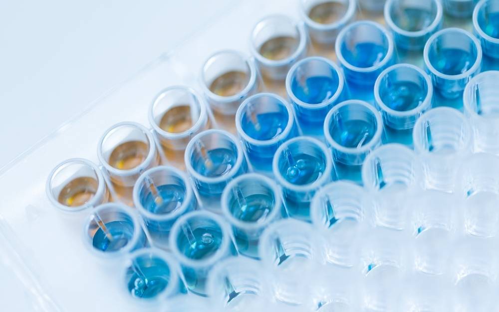
- Detection: A plate reader or imaging system captures the increase in fluorescence, which is directly proportional to the amount of calcium being released.
This sequence allows scientists to quantify the magnitude and the speed of the cellular response. Whether they are looking for a drug that activates a receptor or one that blocks a pathological calcium surge, the assay provides a clear, measurable output that can be analysed and compared across thousands of different samples.
Why this technique is a cornerstone of drug discovery
The pharmaceutical industry relies heavily on the ability to screen vast libraries of chemical compounds to find potential new medicines. A significant portion of modern drugs target a family of proteins known as G-protein coupled receptors (GPCRs). These receptors are involved in almost every physiological process, including sight, smell, and the regulation of mood and immune response. Because many GPCRs signal through calcium pathways, the calcium flux assay has become an indispensable tool for identifying new drug candidates.
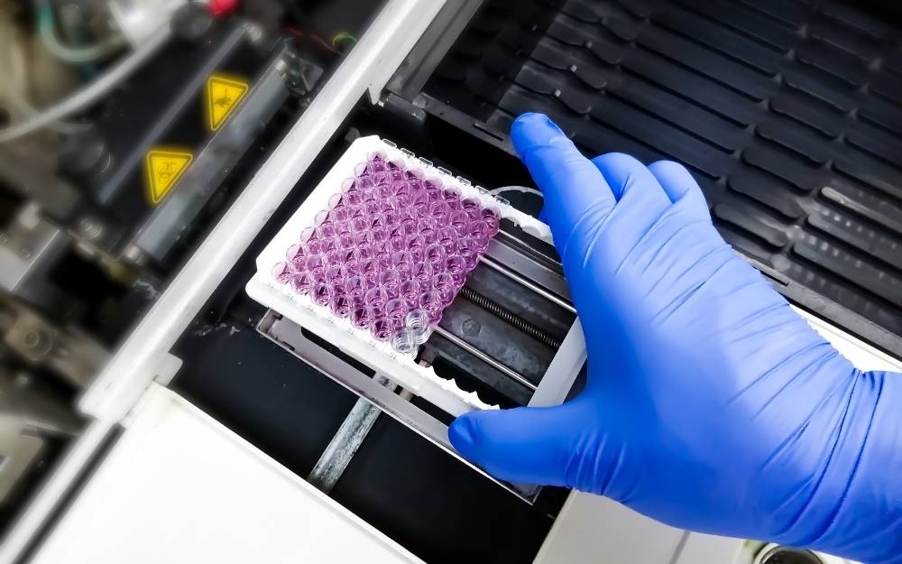
In a high-throughput screening environment, speed and reliability are everything. Modern automated systems can process thousands of samples in a single day, using 384-well or even 1536-well plates. This level of automation allows researchers to rapidly narrow down a list of millions of compounds to a handful of promising ‘hits’. Without the ability to monitor calcium flux efficiently, the development of treatments for conditions like hypertension, chronic pain, and various neurological disorders would be significantly slower and more expensive.
The role of fluorescent indicators in modern labs
The evolution of fluorescent dyes has been a game-changer for this field of study. Early indicators required complex setups and were often difficult to use because they were sensitive to light or leaked out of the cells too quickly. Today, scientists have access to a wide variety of optimised dyes, such as Fluo-4, Fluo-8, and Fura-2, each with its own unique properties.
Some dyes are ‘ratiometric’, meaning they shift their emission or excitation colour depending on whether they are bound to calcium. This is incredibly useful because it helps correct for variations in dye loading or cell thickness, making the data much more robust. Other dyes are designed to be ‘no-wash’, which simplifies the workflow by removing the need to rinse the cells after loading the dye. This reduction in labour-intensive steps not only saves time but also minimises the stress placed on the cells, leading to more natural and reliable biological results.
Optimising the assay for high-throughput screening
While the basic concept of the assay is straightforward, achieving high-quality results in a large-scale screening programme requires careful optimisation. Scientists must balance several factors to ensure they are getting a true representation of cellular activity. If the concentration of the dye is too high, it might actually buffer the calcium and dampen the signal they are trying to measure. If it is too low, the signal-to-noise ratio will be poor, making it difficult to distinguish a real response from background interference.
- Temperature Control: Most cellular processes are highly temperature-dependent. Maintaining a consistent temperature across an entire plate is vital for reproducible data.
- Dispense Speed: The way a drug is added to the wells can impact the result. If the liquid is added too forcefully, it can physically disturb the cells, causing a mechanical calcium spike that masks the drug’s effect.
- Incubation Time: Giving the cells enough time to recover after dye loading and ensuring the dye is properly processed by cellular enzymes is essential for a strong signal.
By carefully controlling these variables, researchers can ensure that their results are a true reflection of the pharmacology they are investigating. This rigorous approach to assay development is what allows for the transition from a simple laboratory observation to a multi-million-pound drug development project.
The impact on safety pharmacology and cardiac research
Beyond the initial discovery of new drugs, the calcium flux assay plays a critical role in safety pharmacology, particularly when it comes to the heart. One of the biggest reasons drugs fail during clinical trials or are withdrawn from the market is cardiotoxicity. If a drug inadvertently interferes with the calcium signalling in heart cells (cardiomyocytes), it can cause dangerous arrhythmias or even heart failure.
By using human-induced pluripotent stem cell-derived cardiomyocytes (hiPSC-CMs), researchers can perform these assays on cells that behave remarkably like a real human heart. Monitoring the calcium handling in these cells allows scientists to see if a potential drug disrupts the rhythmic ‘beating’ of the calcium transients. This early warning system is vital for ensuring that only the safest compounds move forward into human trials, ultimately protecting patients and reducing the risk of late-stage failure in the drug development pipeline.
Overcoming common challenges in data interpretation
Analysing the data from these assays requires a keen eye for detail. A typical calcium flux curve shows a rapid rise followed by a slower decay as the cell works to pump the calcium back out of the cytoplasm. However, not every curve is simple. Some compounds might cause a sustained plateau, while others might trigger a series of oscillations. Interpreting these patterns requires a deep understanding of cellular physiology.
Researchers must also be aware of ‘auto-fluorescence’, where the drug compound itself glows under the same light used to excite the calcium dye. This can lead to false positives if not properly accounted for. Modern software and sophisticated experimental designs, such as using ‘quenchers’ to reduce background noise from the extracellular environment, have made it much easier to filter out these artefacts and focus on the real biological signal. As technology continues to advance, the sensitivity and precision of these measurements will only improve, further cementing the role of this assay in the future of medicine.

Dale is a travel writer with a passion for exploring Japan’s hidden gems. He specializes in cultural travel, local cuisine, and historical landmarks, providing in-depth guides for travelers seeking authentic experiences.